BioExcel’s webinar series continues with a special edition featuring student speakers who were awarded poster prizes at the BioExcel Winter School 2020. Read along to find out more about our speakers and their research.
Date: 18th January, 2021
Time: 15:00 CET
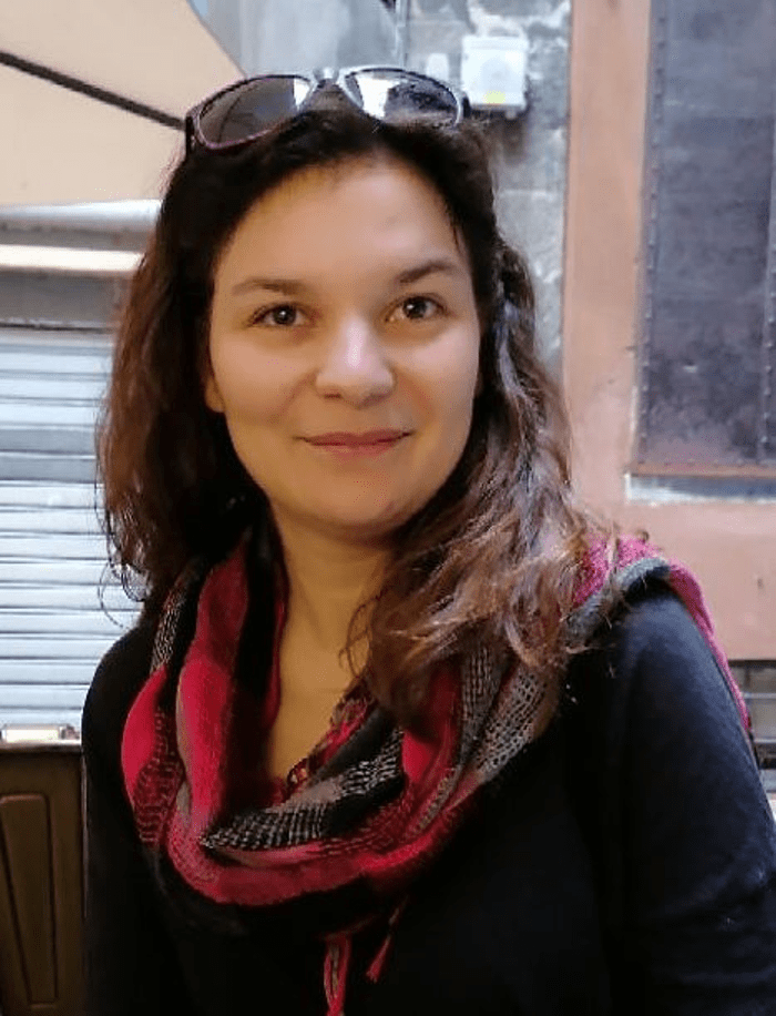
Artemi Bendandi

Artemi Bendandi was born in Thessaloniki, Greece, and studied Physics in the University of Ioannina, Greece. She then pursued a Masters in Physics, in the University of Roma Tre, in Rome, Italy, specialising in Theoretical Physics. Artemi is now concluding her PhD in Physics in the Italian Institute of Technology (IIT) in Genoa, Italy, under the supervision of Dr. Walter Rocchia and Prof. Alberto Diaspro. Her PhD thesis research is in Computational Biophysics, and it concerns the electrostatic interactions in chromatin, the ensemble of proteins and DNA that enables the genome to become compact while remaining accessible. Her primary research covers the computational study of electrostatic interactions between DNA and protein, through the study of the interactions between nucleosome pairs in the Poisson Boltzmann framework, and the examination of the electrostatic importance of the histone tails, the disordered terminal domains of the histones. In her free time she reads science fiction and studies Japanese, and she loves to visit art and history museums.
Modelling Electrostatic interactions and solvation in chromatin: from the single nucleosome to the chromatin fibre
Understanding the mechanisms that trigger chromatin compaction, its patterns, and the factors they depend on, is a fundamental and still open question in Biology. Chromatin compacts and reinforces DNA and is a stable but dynamic structure, to make DNA accessible to proteins. Electrostatic interactions between nucleosomes are central determinants of the topology of chromatin compaction. We present a comprehensive analysis of inter- and intra-nucleosomal electrostatic interactions. We propose a methodology for the study of protein-DNA electrostatic interactions and apply it to clarify the effect of histone tails in nucleosomes. This method can be used to correlate electrostatic interactions to structural and functional features of protein-DNA systems, and can be combined with coarse-grained representations. We focus on the electrostatic field and resulting forces acting on the DNA. We find that the positioning of the histone tails can oppose the attractive pull of the histone core, locally deform the DNA, and tune DNA unwrapping. Small conformational variations in the often overlooked H2A C-terminal tails had significant electrostatic repercussions near the DNA entry and exit sites. The H2A N-terminal tail exerts attractive electrostatic forces towards the histone core in positions where Polymerase II halts its progress. We validate our results with comparisons to previous experimental and computational observations. We use nucleosome pairs at various distances and orientations to numerically study the electrostatic energy and potential in physically viable configurations, using the DelPhi Poisson-Boltzmann solver. We study the contribution of solvation to nucleosome electrostatics and conduct a quantitative analysis on nucleosome porosity.
Roshan Shrestha

I am an M.Sc Physics graduate from Central Department of Physics, Tribhuvan University. My current research focuses on understanding nanoparticle drug delivery mechanisms using molecular dynamics simulations, particularly lipid-nanoparticle interactions and TM-domain protein-nanoparticle interactions in the lipid bilayer.
Twitter: @roshan_shrestha
Molecular Dynamics Simulations of Nanoparticles in Model Bilayers: Lipid Phase Separation and Membrane Protein Interaction
The unique and adjustable properties of nanoparticles present enormous opportunities for their use as targeted drug delivery vectors. For example, nanoparticles functionalized with key surface ligands have been shown to pass through phospholipid bilayers without causing localized disruption. However, biological membranes are a busy and crowded region of a cell and whilst there is evidence to suggest a complex interplay between ligated-nanoparticles with complex biophysical systems, the effect of nanoparticles on the local multi-component phospholipid bilayer environment remains unclear.
The structural stability and downstream signalling potential of integrated transmembrane receptors depend on the interhelical packing of conserved amino acid motifs, such as the Glycine-xxx-Glycine and leucine zipper motifs. An interruption in interhelical transmembrane domain packing can disrupt regular cell function.
In this webinar, we will present our work on the effects that nanoparticles have on lipid phase separation. Using coarse-grained simulations, we observed that bilayer phase separation depends on the degree of unsaturated lipids in a heterogeneous bilayer. In addition, by simulating multi-nanoparticle systems, we show that there is a complex interplay between the lineactant behaviour of the nanoparticles towards the domain interface of the phase-separated regions in the bilayer and its tendency to coagulate in the bilayer to reduce its total exposed surface area. Finally, we also include some recent analysis from several all-atom model simulations of ligated-nanoparticles and the transmembrane domain of the Glycophorin A dimer.
Laura John

During my Bachelor’s and Master’s degree in chemistry, both at the University of Konstanz, Germany, I gained more and more interest in physical chemistry. This led to a Master’s thesis in the lab of Prof Malte Drescher using electron spin resonance spectroscopy for structural and functional analysis of the interaction between the Alzheimer protein tau and the chaperone Hsp90.1–3 After gaining first insights in molecular dynamics simulations, I saw the advantage of combining spectroscopy and simulations for addressing questions in structural biology. Therefore, after graduating in 2017 I made a one-year internship in the computational lab of Prof Mark Sansom at the University of Oxford. After having learned some of the computational techniques I applied successfully for a PhD-position in interdisciplinary bioscience at the DTC, University of Oxford. During my PhD, started in 2018 and supervised by Prof Mark Sansom and Dr Luke Clifton, I combine neutron reflectivity measurements with molecular dynamics simulations to obtain structural and functional information about the interactions between membrane proteins and the lipid bilayer.
Large Scale Membrane Movement Induced by Surface Charge Reversal
Resolving structural and functional aspects of interactions between proteins and membranes requires experiments on membranes which mimic cellular characteristics as closely as possible. In this respect, previous methods which tethered or bound the membrane to a subjacent substrate showed significant deficiency. However, the so-called free-floating bilayer (FFB) model membrane system promises an important step forward. In this FFB system the membrane is surrounded on both sides by water and no longer in contact with the subjacent substrate. This results in better agreement with native membrane dynamics and fluidity. Nevertheless, the acting forces within this system are not fully understood. Ca2+ ions, essential for the self-assembling of the system, seem to play a crucial role. Also, the tightly bound water layers on top of a carboxylate terminated oligo (ethylene glycol) alkanthiol self-assembled monolayer (OEG-SAM), which covers here the substrate surface, are suggested to be a key element for generating FFBs. In this study, molecular dynamics simulations alongside with neutron reflectivity measurements were used to systematically investigate FFB systems consisting of differently charged phospholipid bilayers in presence of Ca2+ and Na+ ions, respectively. The results provided interesting insights in cation- and water-interactions with OEG-SAMs and phospholipid bilayers and enabled to understand the processes within the FFB system. The predicted model suggests that cations bind preferably to the OEG-SAM, hereby attracting the FFB. Nevertheless, structured water layers on top of the OEG-SAM maintain a water layer in between OEG-SAM and FFB via repulsive hydration forces. This knowledge is crucial to use the FFB system for the structural and functional analyses of the interactions between membrane proteins and lipid bilayers.
(1) Weickert, S.; Wawrzyniuk, M.; John, L. H.; Rüdiger, S. G. D.; Drescher, M. The Mechanism of Hsp90-Induced Oligomerizaton of Tau. Sci. Adv. 2020, 6 (11), eaax6999.
(2) John, L.; Drescher, M. Xenopus Laevis Oocytes Preparation for In-Cell EPR Spectroscopy. Bio-protocol 2018, 8 (7), e2798–e2798.
(3) Wojciechowski, F.; Groß, A.; Holder, I. T.; Knörr, L.; Drescher, M.; Hartig, J. S. Pulsed EPR Spectroscopy Distance Measurements of DNA Internally Labelled with Gd 3+-DOTA. Chem. Commun. 2015, 51 (72), 13850–13853.
